Our Dry Eye Clinic offers specialized diagnosis and treatment for dry eye syndrome, providing relief and improving eye health through personalized care.
Ben Franklin is India’s Largest Hospital-Based Optical Chain, operating in over 200 cities across 23 states with more than 400 stores. We are proud to partner with Gitanjali Eye and ENT Hospital to offer international standard optical dispensing to our patients. Our expertise ensures that every patient is offered the right choice of lens and frames to suit their lifestyle, optical requirements, and budget.
Dedicated Healthcare Facility for Eye Speciality in Thiruvananthapuram since 2013
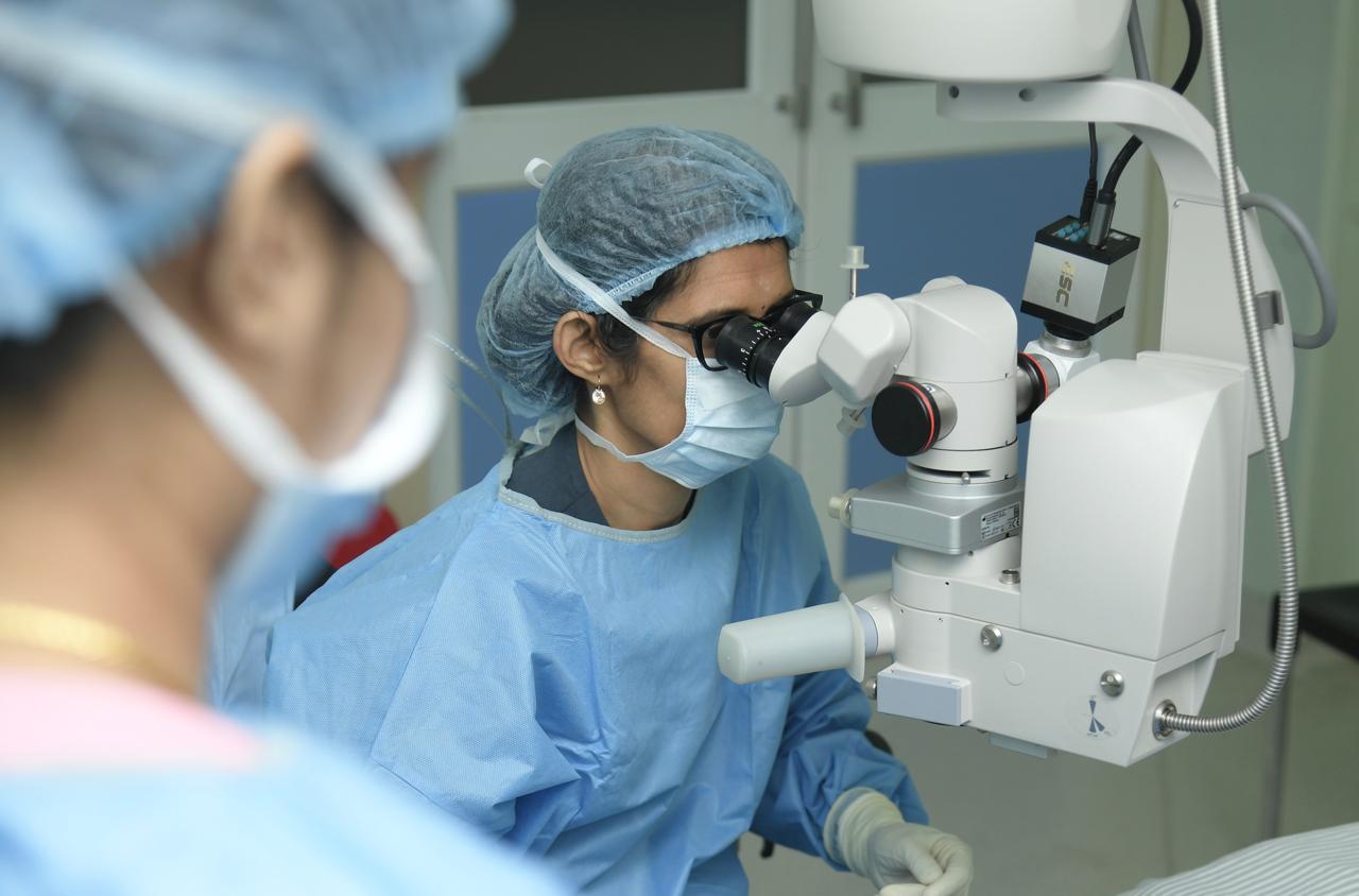
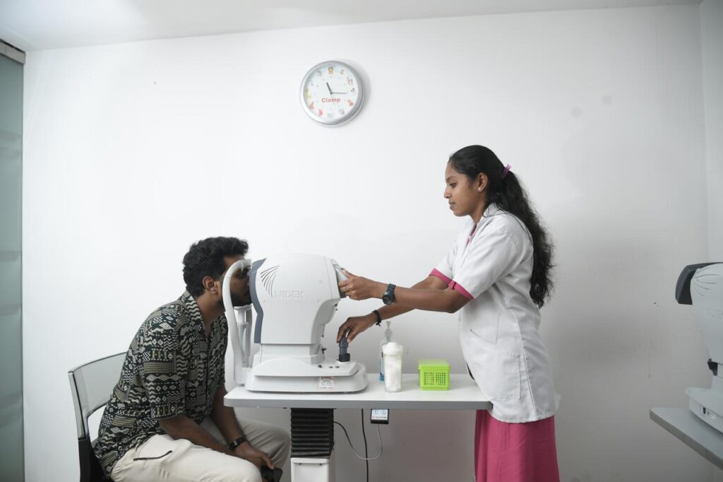
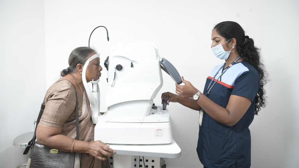
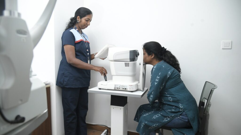
Gitanjali Hospital’s Department of Ophthalmology leverages a state-of-the-art facility to provide comprehensive eye care. Our highly skilled team of ophthalmologists diagnoses and treats a broad spectrum of ocular conditions in both adults and children.
We are dedicated to delivering the highest quality care, supported by:
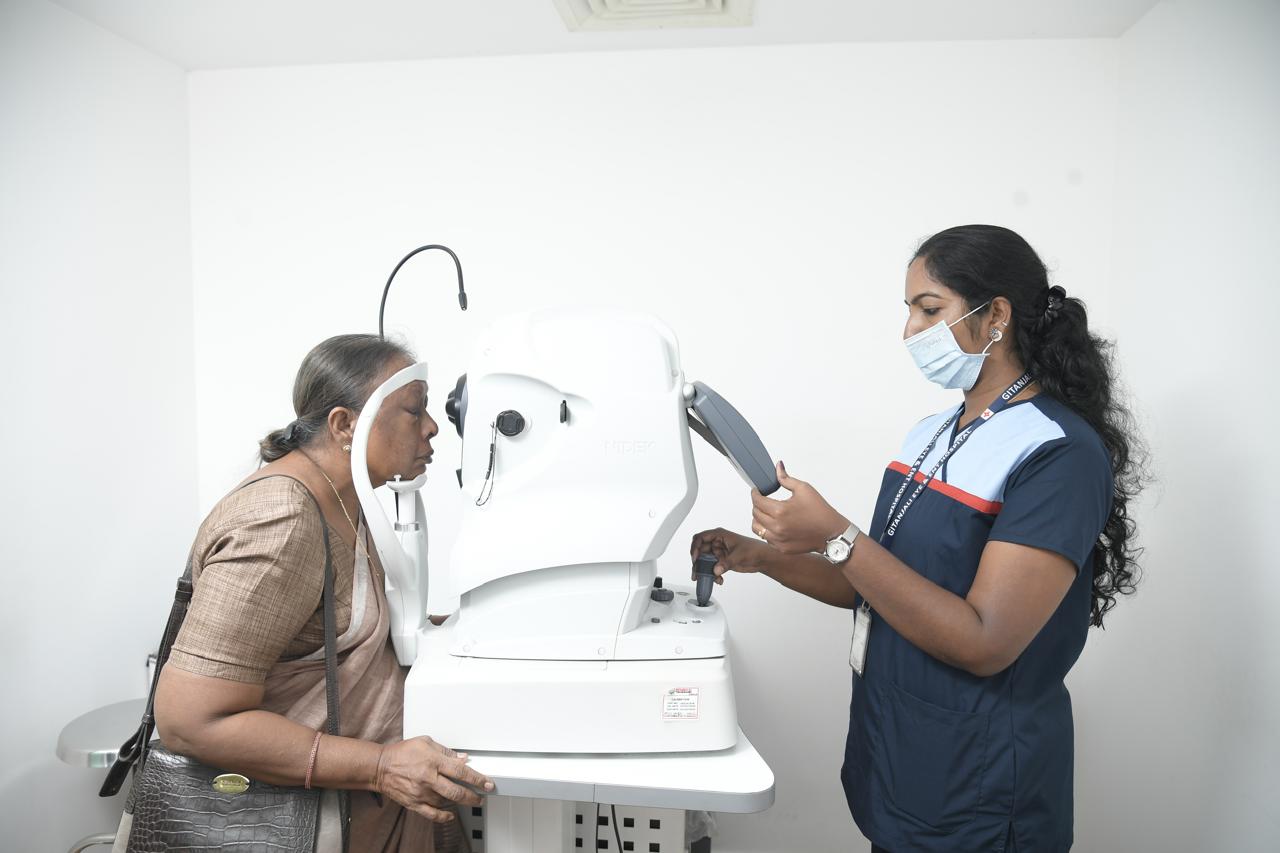
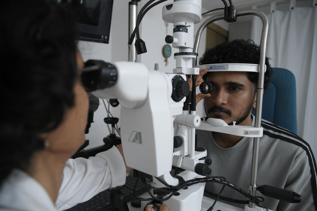
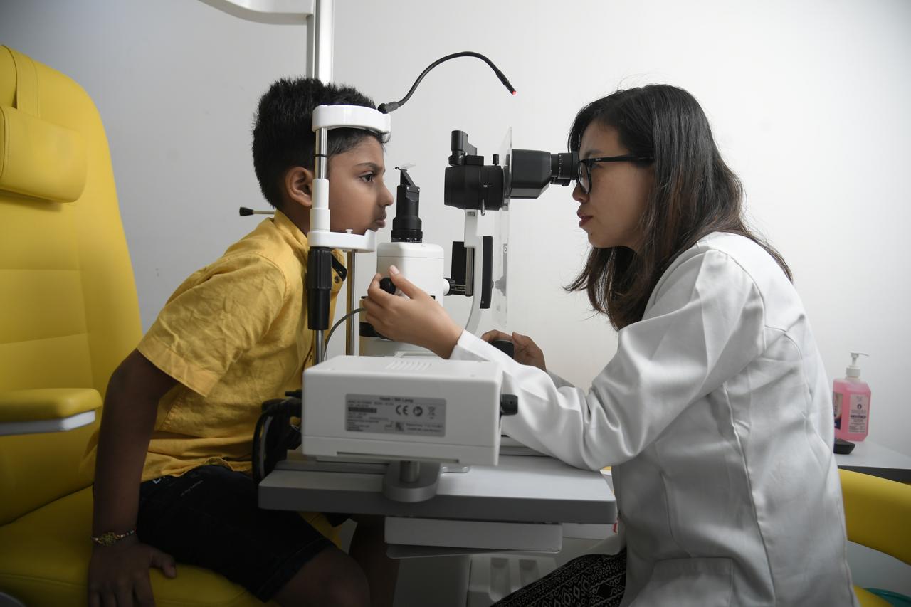
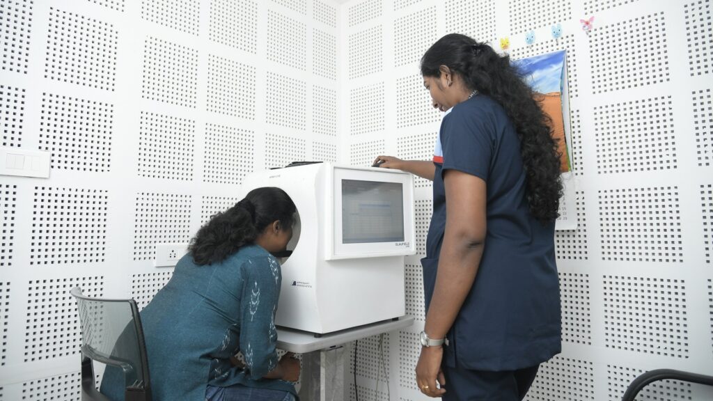
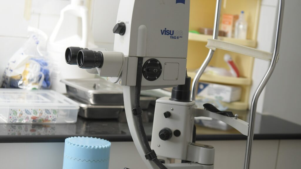
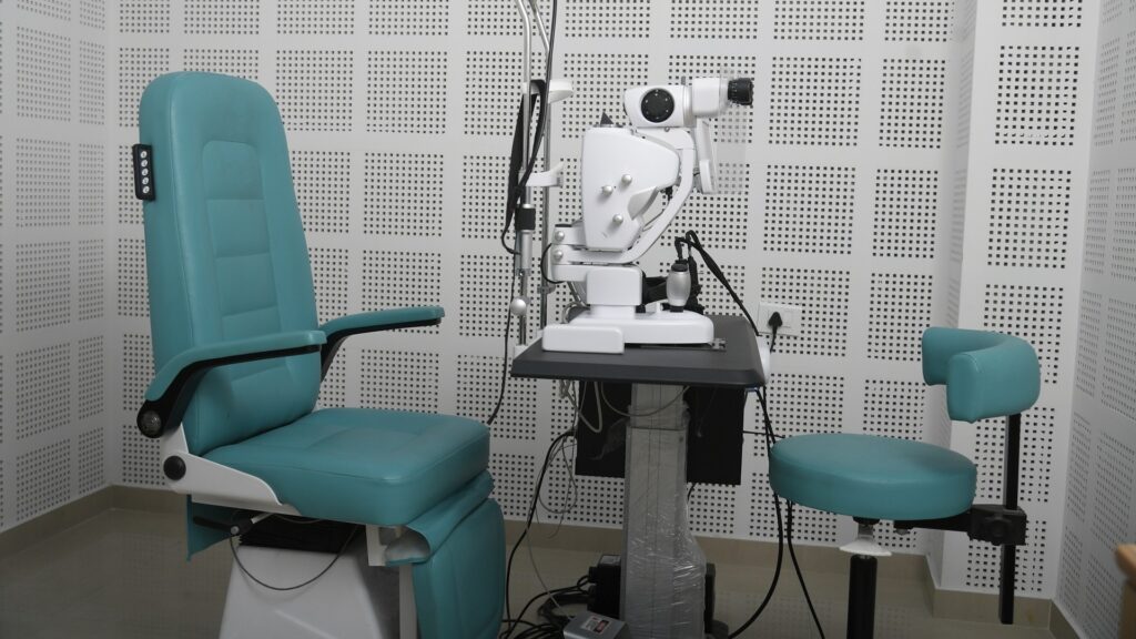
Advanced diagnostic equipment for accurate assessments.
Experienced ophthalmologists for specialized treatment.
Compassionate and dedicated nursing and administrative staff to ensure a positive patient experience.
Our comprehensive services include:
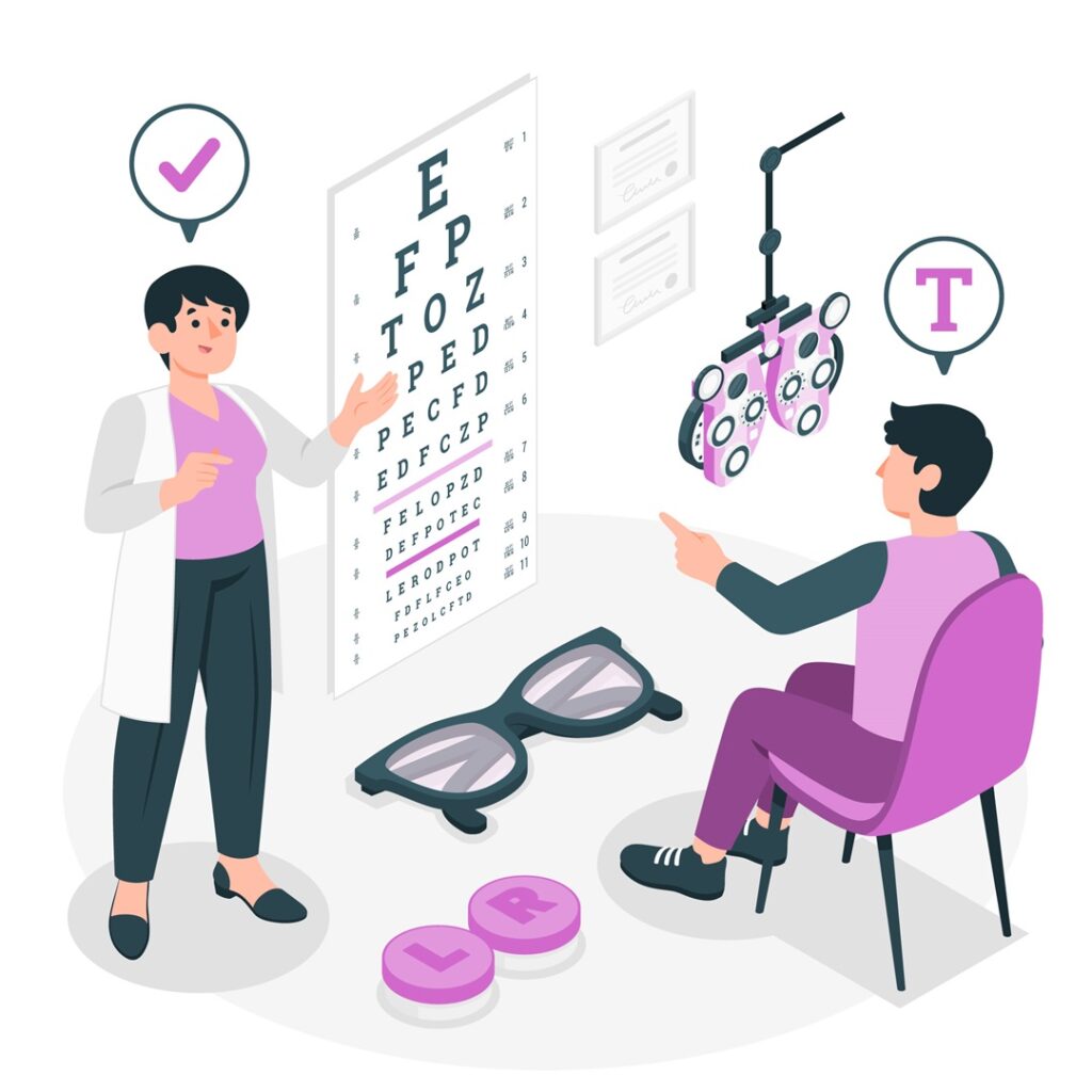
Examinations for routine eye care and specific conditions.
Medical and surgical treatment for various eye diseases.
Pediatric ophthalmology services for children’s eye health.
Meet our Ophthalmologists
Dr Ambika Shetty
DNB, FRCS, FICO, Fellow Phaco & Refractive Surgery
Vitrectomy Vitrectomy is a specialized ophthalmic surgical procedure aimed at addressing a variety of serious eye conditions involving the vitreous body, the …
Our Specialization
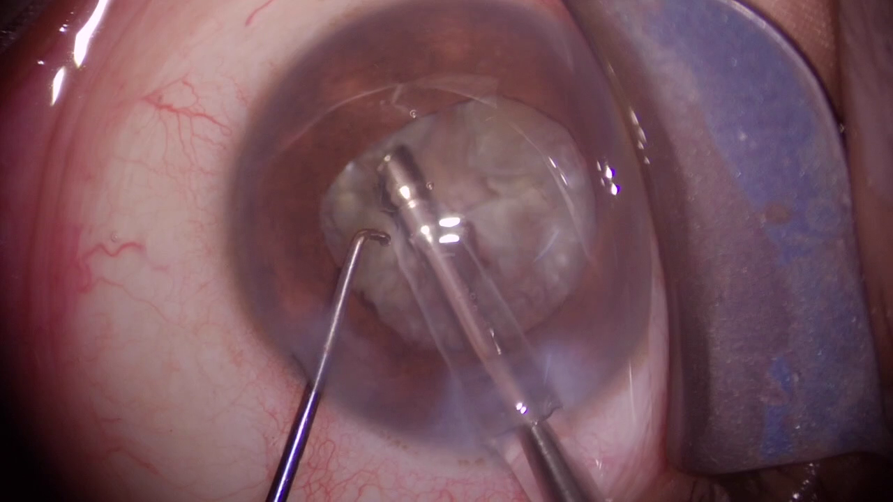
Phacoemulsification Cataract Surgery
The cataract is removed by a surgical technique called phacoemulsification.It is one of the safest surgeries to undergo. However, there are certain situations where the surgeon may explain the risks to you. Complications can occur, but rare. If complications occur such as persistent inflammation, infection etc they must be treated immediately.
The cataract is replaced with a permanent artificial lens implant. This surgery is carried out through a very small incision which reduces healing time and recovery.
The smaller size of the incision is an added advantage to the elderly, diabetic patients, and those with other co-morbidities. This procedure is compatible with most of the available intraocular lenses (IOL), which are placed inside the eye after the removal of the cataract.
Vitreoretinal Surgery
Vitrectomy is a specialized ophthalmic surgical procedure aimed at addressing a variety of serious eye conditions involving the vitreous body, the gel-like substance filling the eye’s interior. Since its inception in the mid-20th century, vitrectomy has evolved into a sophisticated and crucial tool in the field of retinal surgery, offering patients the potential for improved vision and better outcomes in managing complex ocular diseases.
The Anatomy and Function of the Vitreous Body
The vitreous body is a transparent gel-like substance that occupies approximately two-thirds of the eye’s interior space. It is primarily composed of water, collagen fibers, and hyaluronic acid, which help maintain the eye’s shape and provide structural support to the retina. The vitreous body is critical for light transmission through the eye and plays a role in retinal health by keeping the retina in place against the choroid and sclera.
Indications for Vitrectomy
Vitrectomy is indicated in several severe retinal conditions where non-surgical treatments are insufficient. These conditions include:
This occurs when the retina separates from the underlying choroid. Vitrectomy can repair the detachment by removing the vitreous gel, reattaching the retina, and sometimes using a gas bubble or silicone oil to hold the retina in place as it heals.
A macular hole is a defect in the central part of the retina responsible for sharp, central vision. Vitrectomy is performed to remove the vitreous gel pulling on the retina and to encourage closure of the macular hole.
In advanced stages of diabetic retinopathy, the growth of abnormal blood vessels in the retina can lead to bleeding and scarring. Vitrectomy is used to clear out blood and scar tissue to restore vision and prevent further damage.
This condition involves bleeding into the vitreous body, which can obscure vision. Vitrectomy can remove the blood and address the underlying causes of the hemorrhage.
This is a condition where a thin layer of fibrous tissue forms on the retinal surface, distorting vision. Vitrectomy can remove this membrane and improve visual outcomes.
Inflammation of the inner coats of the eye, including the vitreous.
Any intraocular foreign body following trauma.
Dislocated lens/ implants
Vitrectomy remains a cornerstone in the treatment of complex retinal conditions, offering hope for vision restoration in cases where other treatments fail.
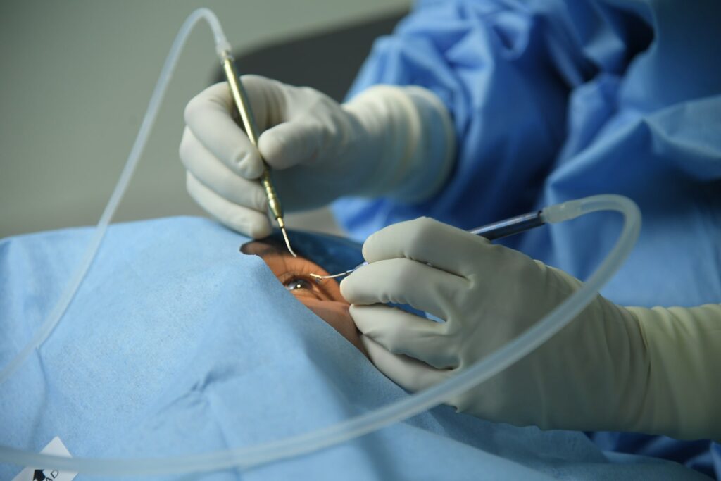
Procedure
The vitrectomy procedure involves several key steps:
The procedure is typically performed under local anesthesia, though general anesthesia may be used in certain cases. The eye is sterilized, and three self sealing incisions are made.
A vitrectomy system with specialized instruments is inserted through tiny incisions in the sclera. The instruments include a vitrector to cut and remove vitreous gel, a light source for visualization, and an endoilluminator.
Depending on the condition, additional procedures such as laser photocoagulation, cryopexy (freezing), or the insertion of a gas bubble or silicone oil may be performed to stabilize the retina
After the surgery, patients may need to maintain a specific head position to ensure the retina heals correctly. Follow-up visits are essential to monitor healing and visual improvement
Surgical Procedures
Phacoemulsification Cataract Surgery
A standard cataract surgery method where the cloudy lens is emulsified and removed using ultrasound technology.
Pterygium Surgery
A procedure to remove abnormal tissue growth on the eye’s surface, restoring comfort and vision.
Mulifocal Intraocular Lens
Advanced lens implants designed to improve vision at multiple distances, reducing dependence on glasses post-surgery
Toric Intraocular Lens
Customized lens implants used to correct astigmatism during cataract surgery for enhanced vision quality
Specialized devices used to expand the pupil during cataract surgery for better access and visibility
External Dacryo cystorhinostomy
A surgical technique to treat tear duct blockages by creating a new drainage pathway for tears.
Lid Surgery
Surgical correction of eyelid abnormalities, such as drooping or excess skin, to improve function and appearance.
Corneal Tear Repair
A procedure to mend a torn cornea, preventing vision loss and restoring the integrity of the eye.
Outpatient Procedures
Automated Visual Field
A computerized test that maps the visual field to detect vision loss due to eye or neurological conditions.
Automated Keratometry
A tool that measures the curvature of the cornea, essential for fitting contact lenses and planning surgeries.
A SCAN Ultrasonography
Ultrasound measurement of the eye’s axial length, used for precise intraocular lens power calculation.
B SCAN Ultrasonography
Imaging technique that provides a detailed view of the eye’s internal structures, especially useful in cases of opaque media.
Chalazion
A small, painless lump on the eyelid caused by a blocked oil gland, often treated with minor surgery.
Lid Trauma Repair
Surgical intervention to correct and heal injuries or damage to the eyelids, restoring function and appearance.
Optical Coherence Tomography
Non-invasive imaging that provides cross-sectional views of the retina, aiding in the diagnosis of retinal and optic nerve diseases.
Noncontact Tonometry
A test to measure intraocular pressure using a puff of air, helping to detect glaucoma.
Nd YAG Laser Capsulotomy / Iridotomy
Laser procedures to treat secondary cataracts or create a hole in the iris to relieve intraocular pressure.
Green laser
A treatment method for retinal conditions, such as diabetic retinopathy, to prevent further vision loss.
Specular Biomicroscopy
A non-invasive imaging technique used to analyze the corneal endothelium, important for assessing corneal health.
Procedures performed
Comprehensive EYE Procedures
Driving License Vision Test
Ensure you meet the vision requirements to get behind the wheel safely.
Gitanjali Hospital offers convenient and comprehensive driving license vision tests. Our experienced eye care professionals will assess your visual acuity, depth perception, and color vision using state-of-the-art equipment.
Invest in your well being
Committed To A Healthier Tomorrow
Let Us Help You Make An Appointment
Skip the waitlist. Book your appointment online and get the care you need, when you need it
Scan the code
Hello there! 👋 Welcome to Gitanjali ENT & Eye Hospital's chat support. We’re here to assist you with any questions you may have, whether it’s about our services, scheduling an appointment, or finding the right specialist.